How does a CT Scan work
CT is a computerised continuous x-ray imaging procedure that generates cross-sectional images or ‘slices’ of the body. Powerful computers use complex software to digitally ‘stack’ these images together to form a 3D image of the patient that we can accurately recognise and interpret. Unlike plain x-ray, a CT scan can differentiate between different types of soft tissue and thus can be used in the investigation of a wide array of conditions. With the administration of an intravenous contrast agent a ‘Contrast CT’ can detect subtle regions of pathology for example in the assessment of metastatic cancers.
What is a CT Scan used for a Eden Veterinary Referrals?
CT scanning is most often used for examining noses, lungs, the contents of the abdomen, and bones. CT has revolutionised the way the veterinary profession looks at problems within complicated joints such as the elbow, for example.
NEW – Outpatient CT Service
At Eden Veterinary Referrals, we want to ensure that every pet has access to the latest in advanced diagnostics, therefore we have launched our Outpatient CT service to all primary care practices.
As veterinary medicine advances, the use of CT imaging becomes increasingly essential not only for the diagnosis of many conditions but also aids with treatment and monitoring protocols
Our CT packages ensure a simple and smooth referral process. If you have any questions about which package is best for your patient, our referral team is on hand to give advice where possible.
Our packages are inclusive of day hospitalisation fees, general anaesthesia, CT scan(s), interpretation report and IVFT as necessary.
Why choose our CT service?
- Affordable, Fast and Reliable
- Hassle free referral service.
- Fixed priced packages.
- Easy online form
- We contact the client.
- Guaranteed appointment with 7 days.
- Imaging report (by specialist) emailed directly to you.
- Direct claims accepted.
- Easy access from M6 J16.
- Separate advice on CT report from a member of our referral team if requested.
- Conversion to full referral based on referring vets’ wishes.
Here are some examples of how useful CT Scans can be…
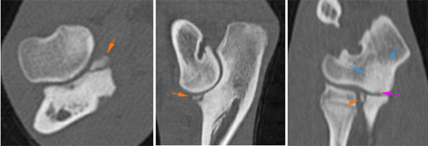
Joints
CT scanning is particularly useful at looking at complex joints (those that are difficult to fully assess with normal X-rays or are composed of more than two bones). CT is used extensively to evaluate the elbow joint, as it is notoriously difficult to assess by radiography.end of antibiotic treatment will also be required prior to surgery.
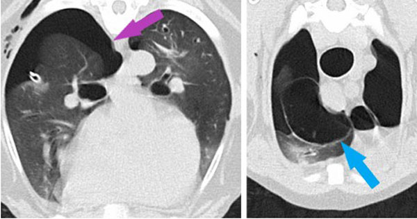
Chest
CT is extremely useful at looking at the chest, particularly for those structures filled with air (the lungs and the windpipe or trachea). It has revolutionised the way we detect diseases that may have been very difficult to assess on normal X-rays. CT is particularly useful at assessing the acute trauma cases we see, allowing us to diagnose the extent of their injuries.
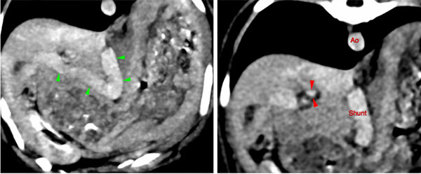
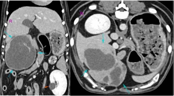
Abdomen
CT scanning is very useful for looking at soft tissues within the abdomen because it is quick (much faster than MRI) and can spot changes that can be seen after contrast (dye) is injected. Using contrast agent is very useful for looking at abnormal blood vessels in the abdomen, such as those seen in a condition called a portosystemic (liver) shunt (see our Portosystemic Shunts Information Sheet), as well as for many other diseases such as neoplasia (cancer).
Nose
CT scanning has largely replaced normal plain radiographs for looking at diseases of the nose. This is because normal X-rays are not very sensitive at looking at nasal disease (that means they often miss the disease that is present) and even worse, can often suggest disease is present when it is not. Nasal cancer is often assessed by CT, in addition to a host of other diseases such as infectious and inflammatory causes of rhinitis, cysts, polyps and foreign bodies to name a few.