What is a Mucocoele?
A salivary mucocoele develops when there is leakage of saliva from the duct into surrounding tissue. Mucocoeles can develop from any of the salivary glands, but the sublingual gland is most commonly affected. The parotid and zygomatic glands are only infrequently affected. There may be a breed predilection (toy dogs, German shepherd dogs) and usually young dogs (1-4 years) are involved. Leaking saliva will follow the path of least resistance, and accumulation may occur anywhere along the length of the duct. Cervical, pharyngeal, and sublingual mucocoeles may be seen. A ranula is another term for a mucocoele, where saliva causes a fluid swelling in the tissues adjacent to the tongue. Pharyngeal mucocoeles may extend some distance into the back of the mouth causing respiratory obstruction. Affected animals may present as an emergency, requiring prompt drainage or temporary marsupialisation.
What causes Mucocoele?
The cause of the injury to the salivary duct is rarely established. Blunt trauma to the head or neck, oral injuries, or sialoliths (salivary stones) have all been suggested as possible causes. However, establishing the cause of the injury is not always important in management of the condition.
How do I know if my dog has Mucocoele?
The most common presentation of sublingual/mandibular mucocoeles is a fluctuant (fluid- filled) swelling at the top of the neck. Often the swelling may intermittently become firm and painful or subside and even regress. In longer standing cases the swelling may migrate towards the midline in which case its origin may become difficult to determine. Less commonly, the mucocoele may develop under the tongue as a ranula and can cause interference with the function of the tongue provoking salivation.
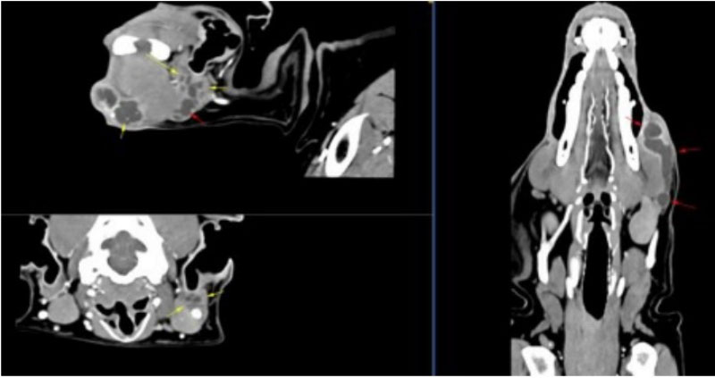
How is Mucocoele Diagnosed?
Mucocoeles in the neck must be differentiated from other swellings (pharyngeal abscesses, lymphomas, thyroid enlargements). Palpation of the mucocoele may determine the glandular origin. Aspiration and identification of the tenacious mucoid blood-tinged fluid is often useful. CT scan are the gold standard imaging modality used to diagnose and fully investigate the cause of a mucocoele.
What Treatments are Available for Mucocoele?
Treatment Options
Drainage of mucocoeles is not a definitive means of management and may even result in the formation of a permanent cervical fistula. Marsupialisation of the mucocoele may be attempted for management of ranula, pharyngeal or zygomatic mucocoeles, though it is rarely successful long term. Removal of the salivary gland is the preferred means of management for mandibular and sublingual mucocoeles. It involves resection of the mandibular and monostomatic portion of the sublingual chain. Although the sublingual gland is the source of the leaking saliva, both the mandibular and sublingual glands on the affected side must be removed. This is because the salivary duct of the mandibular gland is intimately associated with the sublingual gland and cannot be separately resected.
Prognosis
Surgical removal of the salivary gland usually achieves definitive correction of the condition. It is important that all the affected salivary tissue is removed. Recovery from surgery is usually quick, but it can take several days for the large swelling to subside after surgery. An indwelling drain may be required following surgery while the tissues heal. For large mucocoeles, a dog may have to stay in hospital for several days while the wound settles down. Once the wound has healed, however, dogs quickly return to normal activities They have more than enough salivary tissue to make up for any loss. If you have any further questions about salivary mucocoeles you should speak to your veterinary surgeon who will be able to discuss this condition with you more fully.