What is Urinary Incontinence?
Urinary incontinence is defined as the involuntary leakage of urine, and it occurs mainly when a dog/cat is most relaxed. There are several causes for urine leakage:
- Overflow problems when the bladder is so full that the valve mechanisms cannot prevent leakage
- Urinary tract infection/irritation, inflammation of the bladder, prostate or vagina, and growths can all stimulate increased frequency of urination
- Increased water intake which can lead to increased urination
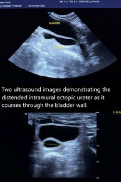
- Anatomical abnormalities can predispose urine leakage, the most common form of which is incorrect entry of the ureters into the bladder (ectopic ureter). Ectopic ureters usually open at the neck of the bladder or into the urethra and this cause urinary incontinence.
Approximately 80% of mature dogs referred for investigation of urinary incontinence are diagnosed with urethral sphincter mechanism incompetence (USMI). USMI can be congenital (present at birth) or acquired and can occur together with ectopic ureters, with the result that cases with ectopic ureters (which are present from birth) can sometimes present later in life as the valve mechanism also gradually ‘gives up’.
What Animals are Most Commonly Affected by Urinary Incontinence?
Urinary incontinence is most common in middle aged to older medium large female dogs. Male dogs are affected less due to the greater length of the urethra. Cats are rarely affected.
Neutered female dogs are more likely to become incontinent, and those that do often do so within one or two years of neutering. The timing of a neutering seems to have some impact on this disease.
Most surgeons prefer to neuter female dogs after one or two seasons, particularly in breeds with a predisposition for incontinence.
How is Urinary Incontinence Diagnosed?
Whilst the majority of dogs who develop incontinence in later life suffer from USMI, it is important to rule out any factors that may contribute to incontinence before considering treatment for USMI. The diagnosis and treatment of any coexisting abnormality may significantly increase the likelihood of successful treatment of or even completely avoid the need for specific treatment for USMI.
Investigation of the entire urinary tract is extremely important as there are no specific tests available to diagnose USMI. As such the diagnosis is reached by rolling in or out the underlying problems.
- Blood tests: to check for evidence of kidney disease or hormonal imbalances
- Urine analysis: Urinary infections are common in incontinent patients and can cause complications if not diagnosed and treated before surgery
- Ultrasound scan: to examine the kidneys and bladder for abnormalities and for evidence of where the ureters enter into the bladder
- Intravenous Urogram (IVU); a contrast which shows up on radiographs is injected into the bloodstream and followed into the kidneys, ureters and bladder. This is useful to detect abnormalities of the ureters, including ectopic ureters. A CT is often preferred.
- Double contrast cystogram; air and dye are injected up the urethra into the bladder. This is useful for examining the position of the bladder neck and when looking for bladder wall abnormalities or evidence of bladder stones
- Retrograde studies: dye is injected into the vagina, the urethra and bladder to examine the anatomy of the vagina and possibly reveal ectopic ureters not seen in the IVU
- Cystoscopy: a small camera can be placed in the bladder neck via the urethra in medium and large breed dogs. This technique is very useful in ruling out ectopic ureters.
If a urine infection is diagnosed, this will require antibiotic treatment for four to six weeks. A negative culture result following the end of antibiotic treatment will also be required prior to surgery.
What Treatments are Available for Urinary Incontinence?
Where USMI is the only or major cause of urine leakage a good response can be achieved through medical therapy.
Surgery for uncomplicated USMI is reserved for dogs where medical treatment is unsuccessful, or continuous therapy proves impractical. There are several techniques available to increase the resistance to leakage of urine, all of which have very similar success rates.
- Urethropexy (males and females); The urethra is surgically fixed in a position to improve continence
- Colposuspension (females only); The vagina, associated urethra, and bladder neck, are surgically held in a position to improve continence
- Collagen injections ; into the bladder neck via a camera placed in the urethra (males require a modification of this technique). Injections need to be repeated after two to three years
- Prostatopexy (males only); the prostate gland is used as an anchoring point to improve the positioning of the urethra and bladder neck.
- Ectopic Ureter Surgery – correction of the opening location of the ureters either by an open surgical approach or by a minimally invasive approach using the cystoscope.
What can I Expect if my Pet is Treated for Urinary Incontinence?
Research suggests that the outlook following surgery is the same regardless of the technique used:
50% -60% become continent
30% – 40% are improved but require some supplemental medication to remain continent
10% – 20% do not benefit from surgery.
Complications of the different surgical procedures are similar regardless of technique used. These include infection, urine retention and continued incontinence. The choice of surgical technique used will be tailored to the individual pet and based on the evidence gained during the investigation and the discussions between the veterinary surgeon and the owner.