MRI Scanning
Founded in 2003, Burgess Diagnostics provides an innovative and pioneering mobile imaging service for the veterinary sector. Burgess MRI comes to Eden Vets on a minimum of a monthly basis. All of their highly qualified clinical staff are specialist radiographers who have each held senior clinical positions for several years. With cutting-edge equipment, a wealth of experience and expertise in all aspects of MR Imaging, we can provide a high-quality scanning service which delivers images of the highest resolution.
How does a MRI work
Magnetic resonance imaging (MRI) is a type of scan that uses a strong, static magnetic fields and radio waves to produce detailed images of the inside of the body by aligning the usually randomly orientated atomic nuclei of hydrogen atoms in the patient’s body.
A radio frequency (RF) pulse is then applied, forcing the spinning hydrogen protons to wobble. Once the RF pulse is turned off, the wobbling of the hydrogen protons generates a small electrical signal.
The signal is detected by RF coils, which need to be positioned as close to the anatomical area of interest as possible.
The rate at which the protons return to their equilibrium state distinguishes different bodily tissues.
A powerful computer then analyses the signals that are received by the coils, and this builds up a picture of the parts of the body which are being investigated. Sophisticated changes can be made to the magnetic field and radio frequencies to highlight different tissues and different diseases within the tissues.
Which cases would we choose MRI for?
- MRI can image through bony structures and so is the treatment of choice for images of the spine, spinal cord and brain disorders.
- Brain tumours
- Lumbosacral disease
- Tendon and ligament injuries
- Investigation of seizures
- Vestibular problems
- Neck and back pain
- Provides excellent detail for pre-operative planning for many surgical conditions.
MRI Package Prices
£2000 for 1 area
£2500 for 2-3 areas
This fee includes contrast, standard interpretation, return pet ambulance transport, to & from the MRI centre
Here are some examples of how MRI is Useful
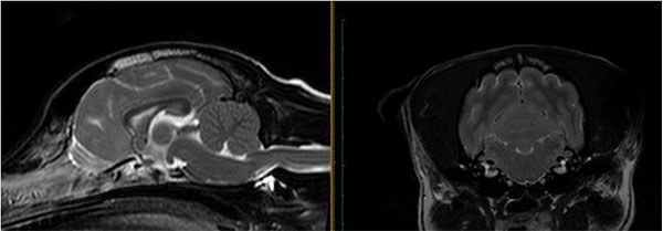
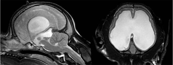
Brain
As the brain is surrounded by a hard bony case (the skull), other types of imaging such as X-rays struggle to produce useful images of the brain tissue. MRI is gold standard for gaining intricate detail of the brain and the structures surrounding it. CT scanning can also be used, however the images are not as clear when compared to MRI therefore lesions may be missed.


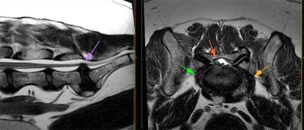
Lumbosacral Disease
MRI is the best method for confirming the presence of many spinal conditions, including lumbosacral disease. It provides detailed information on the location and extent of degenerative and compressive changes to the intervertebral disc, spinal cord and nerves in the lumbosacral junction

Spine
MRI provides superb detail of the spinal cord and surrounding soft tissues in a safe, non-invasive fashion and is classed as ‘gold standard’ for imaging the spine. Some examples of conditions MRI would diagnose are as follows:
- IVDD – Intervertebral disc disease (type I, II and III)
- FCE – Fibrocartilaginous Embolism
- Neoplasia (cancers) of the spinal cord and associated structures
- Spinal Cord Contusion injuries
- Nerve root conditions
A CT scan can also be used to successfully image the spinal cord in emergency trauma and certain types of IVDD cases. However, CT does not provide us with the detailed information when compared to MRI for those more complex, chronic, or progressive cases. In some instances, both imaging modalities can be used in combination to gain a complete picture of these structures.

