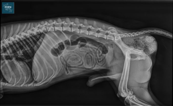
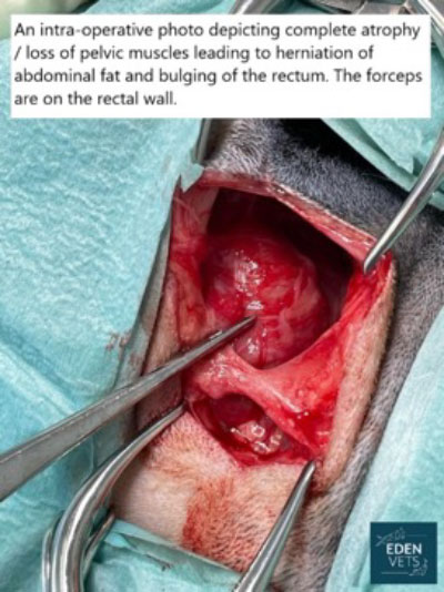
What is a Perineal Hernia?
A perineal Hernia (rupture) is characterised by progressive weakening of the muscles of the pelvic diaphragm. It occurs almost exclusively in the male dog, although rare cases are reported in the bitch, and the cat. The precise cause of the condition remains obscure but is believed to be associated with hormonal influences on the pelvic diaphragm, straining, and congenital or acquired weakness.
The condition develops following deterioration in the supporting function of the pelvic diaphragm. In the normal animal, the pelvic diaphragm provides lateral support to the rectum as it exits the pelvic canal. Disruption to this structure permits an increase in rectal capacity, and interference to the normal linear passage of stool during defecation. Rectal enlargement, faecal impaction and straining to pass a motion are the inevitable consequences. In severe cases the bladder can also become trapped within the hernia which can cause an inability to urinate leading to a life-threatening surgical condition.
Clinical signs can include:
- Perineal swelling – this can fluctuate in size.
- Perineal bruising
- Pain/discomfort associated with the swelling.
- Pain or discomfort when passing of faeces.
- Constipation
- Urine retention – an inability to empty the bladder properly.
- Acute illness due to obstruction of urine flow from the bladder
- Generalised malaise
What investigations are involved to diagnose this condition?
A reducible swelling may be present in most cases. To confirm the diagnosis, rectal examination will be required to reveal a defect in the pelvic diaphragm. In some animals, an abnormal size of the rectum may be apparent.
Ultrasonographic examination of the rupture and/or contrast x-rays will permit localisation of the prostate and bladder. Urine and blood tests may be required along with chest x-rays in some cases.

How is this condition treated?
Perineal Hernia is a surgical condition, and there is usually little benefit in delaying repair. In fact, early repair greatly reduces the risk of post-operative complications such as incontinence (due to progressive stretching of the perineal wall).
Those cases with other disease processes that have contributed to the development of the rupture will need to receive treatment for that underlying problem, prior to or at the same time as repair of the rupture.
Some ruptures are repaired using the muscles that are present within the pelvic diaphragm, although many require movement of additional muscle into the area to close the rupture. In some severe cases, mobilisation of a muscle from a hind leg or a synthetic mesh may be required to complete the closure.
Further support for the repair can be given by fixing the bladder and colon (large intestine) into the abdomen but this is not necessary in all cases.
Perineal rupture repair is straightforward in experienced hands, and recurrence rates of less than 10% are reported.

What is the prognosis?
Over 90% of uncomplicated cases receiving rupture repair and castration resolve following the surgery. In experienced hands, complications following surgery are infrequent, and animals usually stay in the hospital for a few days to ensure they are comfortable and passing faeces easily.
Despite careful selection of the technique used and meticulous care, complications can occur in a minority of cases, and these include:
- Recurrence of the rupture – stool softeners may be advised long term to reduce the straining during defaecation and therefore protect the repair.
- Herniation of the other side – as most cases are bilateral (affecting both sides).
- Infection (the surgical site is adjacent to the anus which is a source of bacteria)
- Faecal incontinence – this is uncommon and usually temporary.
