What is a total Ear Canal Ablation Surgery (TECA-BO)?
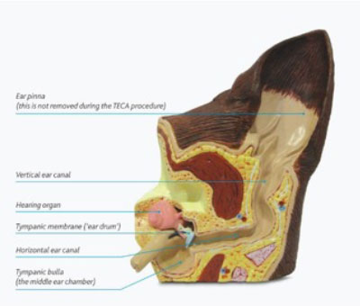
The ear is composed of the outer flap (the pinna), the canal leading to the ear drum, the middle ear (the bulla) and the inner ear (including the tiny bones of the ear and the balance control).
Why perform a TECA-BO?
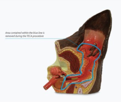
TECA-BO is often performed following failed medical treatment of external or middle ear disease. It is a salvage procedure, meaning that removal of the entire canal cannot be undone, and will result in deafness in that ear. It involves the removal of the diseased and infected ear canal whilst leaving the inner ear (the hearing organ) itself (the inner ear) in place. The middle ear chamber (tympanic bulla) is carefully inspected, and any abnormal tissue or material is removed.
What Animals are Most Commonly Affected by Urinary Incontinence?
Urinary incontinence is most common in middle aged to older medium large female dogs. Male dogs are affected less due to the greater length of the urethra. Cats are rarely affected.
Neutered female dogs are more likely to become incontinent, and those that do often do so within one or two years of neutering. The timing of a neutering seems to have some impact on this disease.
Most surgeons prefer to neuter female dogs after one or two seasons, particularly in breeds with a predisposition for incontinence.
Investigation and Surgery
A member of our surgical team will discuss your pet’s history and symptoms to decide a tailor-made diagnostic plan. This will include a full physical examination, including an ear examination (which is sometimes easier once your pet is asleep), and often includes a CT-scan or an MRI of the head. This will determine the level of disease and which surgery may be most appropriate. Surgery is typically performed a day later to diagnostics, so that a complete plan can be made. The flap of the ear is not removed, but the ear canal is removed in its entirety, including the ear drum, and the lining of the middle ear.
When performed by a Specialist Surgeon, more than 90% of owners report a significant improvement in their dog’s quality of life following the procedure. Whilst most procedures result in success, there are some potential complications that can occur. Our Specialist Surgeons will take every step possible to minimise the risk of complications and maximise the chances of a good outcome.


Possible complications include:
- Reduced hearing sensitivity; similar to the effect of ear plugs or being under water
- Facial nerve paralysis; can occur 10-20% of cases and is usually temporary. The nerve runs very close to the ear canal and can be bruised during the procedure. Paralysis results in some mild drooping of the upper lip and eyelid on the affected side and can result in a reduced blink reflex. Eye lubrication may be required for a time until the third eyelid adapts. Most cases resolve over 4 to 6 weeks.
- Bleeding; significant bleeding can occur during the TECA operation due to the location of blood vessels. All possible steps will be taken to avoid bleeding and minimise the risk of life threatening levels of haemorrhage.
- Wound healing complications; patients will need to wear an Elizabethan collar following the procedure until the stitches are removed.
- Vestibular syndrome (balance problems); the organ responsible for balance is positioned next to the organ responsible for hearing. In some rare cases damage to this region is unavoidable due to the extent of the disease process. Dogs with the complication of balance dysfunction (similar to vertigo) often require significantly more nursing in the immediate postoperative period and some may never fully recover.
- Infection/abscess formation; when performed by Specialist Surgeons the occurrence infection or abscesses are is very low. Operating in such infected tissue can result in the risk of abscessation which can prove extremely difficult to resolve. Further surgery may be required and not all cases are successfully resolved.
- Horner’s syndrome; a combination of clinical signs that can result from damage to a complex of nerves that cross the middle ear. Damage to these nerves causes protrusion of the third eyelid, drooping of the upper eyelid and constriction of the pupil of the eye on the operated side. These signs are infrequent in the dog, and are usually temporary. Those cases that do not fully recover do not experience a significant reduction in quality of life or ability to see
Why have I been referred?
TECA is complex surgery, which should be performed once other options have been exhausted. Thorough knowledge of the complex anatomy of the head is needed to avoid surgical complications. Advanced diagnostic facilities aid in identifying the underlying cause, which can affect surgical planning purposes. The surgery is performed by one of our experienced surgeons, who will explain the pros and cons of each option. Your pet will be hospitalised for individually tailored pain management, and our hospital is staffed 24h per day with vets and nurses, who can assist your pet’s recovery from surgery and anaesthesia.
Prognosis
Most dogs do very well after TECA, and clients often report a significant improvement in their pet’s quality of life. Unfortunately, the surgery does come with some risk of complications as stated above therefore clients need to be fully appreciative and accepting of this procedure prior to going ahead.
