PATIENT: Amber, 3yr 2m old FN Toy Poodle
Amber, a 3-year-old FN toy poodle presented as an emergency referral to Eden Vets in a comatose condition. She had been behaving abnormally for a few weeks with the owners reporting she was circling the perimeter of the room and only turning when bumping into the wall.
They reported that her appetite had been decreased and was PU/PD. Her pulses were weak but palpable with a rate of 160bpm. Cardiac auscultation revealed no murmurs nor arrythmias. Systolic blood pressure measured with a Doppler was 90mmHg indicating hypotension. Abdominal palpation was unremarkable, and temperature was normal.
Bloods were carried out which indicated severe dehydration (hypernatremia, hyperchloremia and haemoconcentration (HCT 68%)). There was a mild stress pattern leucogram and mild increases in ALT and Phos but otherwise bloods were unremarkable. Basal cortisol was 312nmol/L, excluding hypoadrenocorticism. Resting bile acids were mildly elevated at 19.7umol/L. CRP was markedly elevated at 164 indicating acute inflammation. A urine sample was obtained via cystocentesis. USG was concentrated at 1.035 but less then would be expected for the degree of dehydration. The urine
was grossly brown and on cytology there was pyuria with visible cocci and rods and profuse urate crystalluria.
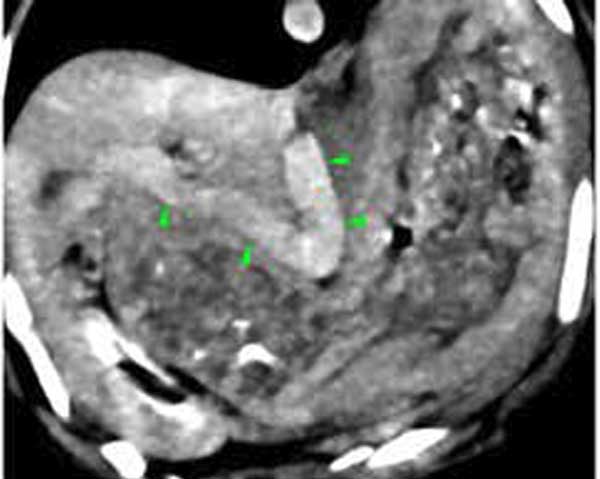
Abdominal ultrasound and contrast CT scan confirmed a single large extra hepatic portosystemic shunt measuring 0.5cm in diameter with associated reduced hepatic volume and portal hypoplasia The medical team stabilised Amber using a combination of medical and dietary management and several weeks later was presented for surgery. Contrast CT scan of brain revealed no abnormalities.
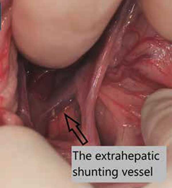
A hepatic sparing general anaesthesia protocol was utilized and the patient clipped and aseptically prepared. A moderately sized ventral midline celiotomy was performed to allow for adequate abdominal inspection. The EHPSS was located and given the large diameter of the shunt with associated portal hypoplasia, a 6mm Ameroid constrictor was placed for slow, gradual attenuation. The surgery went well, and the patient recovered uneventfully.
Amber remained hospitalised for 7 days post-operatively and after developing mild ascites (believed to caused by the weight of the ameroid pressing on the shunt) was successfully discharged. No other complications were seen. At her 6-month re-check, Amber was doing well and is currently being weaned off medical management.