PATIENT: Rita, 3yr 6m old FE Hungarian Vizsla
Rita presented to the orthopaedic department with severe episodic right pelvic limb lameness. The owner described that she would intermittently scream and hold the right pelvic limb high in the air
In addition, she would offload the limb when standing still and was less keen to exercise to the same levels as before. The symptoms were non-responsive to standard analgesic medication and Rita was referred for further assessment
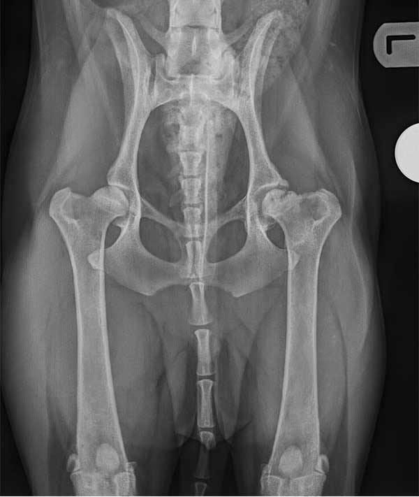
Examination localized the pain to the lumbosacral spine. Rita was painful on full hip extension, direct palpation of the LS region, tail jack test and when palpating the sciatic tract. Referring vets’ radiographs were suggestive of a lumbosacral vertebral malformation and it was advised that Rita should have both MRI and CT to fully assess the neural and osseous structures in the LS region.
The advanced imaging findings were:
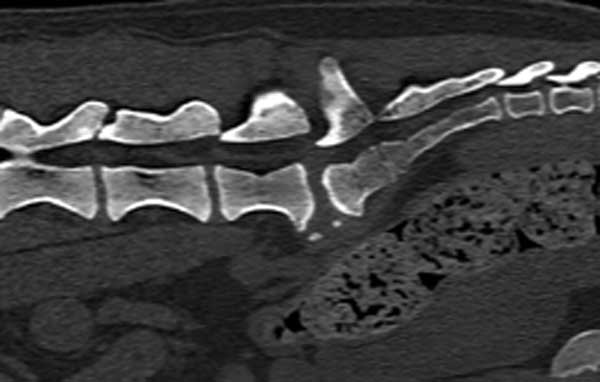
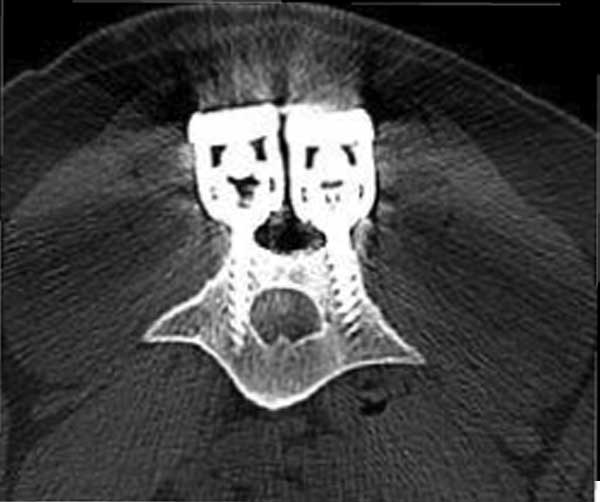
1. Lumbarised sacrum (Transitional vertebra)
2. Retrolisthesis of the sacrum with respect to L7
3. Mild protrusion of the LS disc
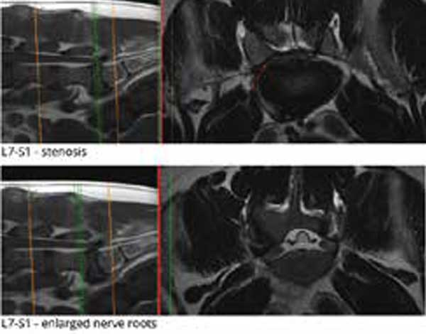
4. Significant stenosis of the right neuroforamen – both nerve roots at the level of the LS disc were enlarged consistent with reactive neuritis.
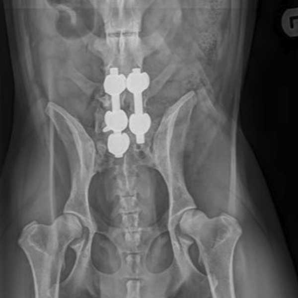
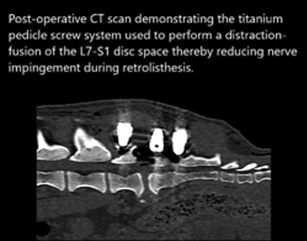
The degree of foraminal stenosis and retrolisthesis were the cause of the severe right pelvic limb lameness and warranted surgical treatment. A dorsal laminectomy to remove the sacral bone that was causing nerve impingement during retrolisthesis was performed followed by a distraction-fusion of the L7-S1 disc space using a titanium pedicle screw system. Distraction of the affected vertebral segments reduces neural compression by increasing the size of the neuroforamen through which the nerve root travels, the subsequent fusion fixes the vertebral segments in place and prevents ongoing dynamic compression.
Rita made an excellent recovery and is now back to normal exercise without restriction.